Body Scan Detects Skin Cancer
December 9, 2001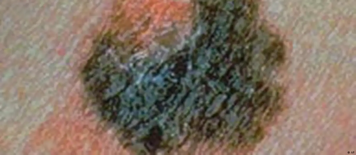
Skin cancer is on the rise world wide. The incidence has increased 20-fold since the 1930s. In Germany, alone some 100,000 people are diagnosed with malignant melanomas every year. According to experts, within the next ten years, the number of people with skin cancer will double.
The rapid increase in skin cancer is alarming. But, compared to other forms of cancer, it is relatively easy to detect and treat if caught early enough. Unfortunately though, many people often ignore or are unaware of the early warning signs.
Scientists at the Fraunhof Institute for Biomedical Technology in St. Ingebert have developed a new form of software to assist dermatologists in the early detection of malignant melanomas.
The doctor takes photographs of a patient using either a digital camera or a video camera. When the patient comes for a second check up, the dermatologist takes a second set of pictures and feeds them into the new software program, which compares and analyses the two sets of pictures.
The software is programmed to recognize changes in the skin’s surface. It can indicate whether or not new moles have appeared or their pigmentation and structure have changed. The software blends the two sets of pictures and presents the doctor with a "before and after" image on the monitor. Critical changes in the skin are highlighted with different colors or blinking circles.
When the scientists developed the software they had to take two key factors into consideration. First, in the time between the first and second set of photos, a person’s skin naturally changes. Second, it is nearly impossible for the doctor to photograph the patient two times in exactly the same position with exactly the same light.
"For this reason, the software has to automatically correct the pictures to account for such differences before they can be compared with one another," said Dr. Frank Volke, director of the research group Magnetic Resonance. Such automatic corrections are made possible with a series of complex mathematical algorithms, which were also developed at the Fraunhof Institute.